Home » Without Label » Upper Leg Tendon Anatomy / PPT - The Knee Joint PowerPoint Presentation - ID:634195 / Three vastus muscles and the rectus femoris.
Upper Leg Tendon Anatomy / PPT - The Knee Joint PowerPoint Presentation - ID:634195 / Three vastus muscles and the rectus femoris.
Upper Leg Tendon Anatomy / PPT - The Knee Joint PowerPoint Presentation - ID:634195 / Three vastus muscles and the rectus femoris.. The quadriceps and hamstring muscles work together to straighten (extend) and bend (flex) the leg. The muscles of the anterior thigh consist of the quadriceps (or quads): This important tendon in the back of the calf and ankle stores the elastic energy needed for running, jumping, and other physical activity. Another large hip flexor is the rectus femoris. The hamstring muscles in the back of the thigh, the quadriceps muscles in the front, and the adductor (groin) muscles on the inside.
This deep muscle begins in the low back and pelvis and connects on the inside edge of the upper femur. The quadriceps tendon attaches the quadriceps muscles to the patella. For a more detailed anatomy of the muscle, check out the following leg muscle diagrams posted below. The largest muscle masses in the leg are present in the thigh and the calf. This important tendon in the back of the calf and ankle stores the elastic energy needed for running, jumping, and other physical activity.
Pin by Paul Neale on Anatomy | Pinterest | Legs, Search ... from s-media-cache-ak0.pinimg.com It is also visible on the medial edge of the thigh from the anterior. The legs are the lower limbs of the human body that provide support and stability in addition to allowing movement. It is the junction of the thigh and the leg and is a hinge joint. This deep muscle begins in the low back and pelvis and connects on the inside edge of the upper femur. The human leg, in the general word sense, is the entire lower limb of the human body, including the foot, thigh and even the hip or gluteal region. The posterior upper leg muscles provide your knees with mobility (extension, flexion and rotation) and strength.they work closely with your quadriceps muscles at the front of your thigh, your gluteal muscles, and your calf muscles to ensure proper movement of your leg and hip. It arises by a thin aponeurosis from the anterior margins of the lower half of the symphysis pubis and the upper half of the pubic arch. The muscles of the anterior thigh consist of the quadriceps (or quads):
This mri wrist coronal cross sectional anatomy tool is absolutely free to use.
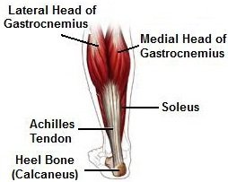
Where are the tendons in your upper legs : Upper leg pain may be the result of systemic disease, such as the following. For a more detailed anatomy of the muscle, check out the following leg muscle diagrams posted below. Legs are used for standing, and all forms of. Your lower leg includes three main muscles, located behind your tibia or shinbone. Anatomy the four quadriceps muscles meet just above the kneecap (patella) to form the quadriceps tendon. The legs include the upper leg, knee, lower leg, ankle, and. The four muscles all extend the lower leg. It is also visible on the medial edge of the thigh from the anterior. The main functions of the quads are flexion (bending) of the hip and extension (straightening) of the knee. Vastus medialis, intermedius, lateralis and rectus femoris muscles. The single bone in the thigh region is called the femur. The calf muscles are pivotal to movement of the ankle, foot, and toes.
They consist of the rectus femoris, vastus intermedius, vastus lateralis and the vastus medialis. The legs include the upper leg, knee, lower leg, ankle, and. The largest muscle masses in the leg are present in the thigh and the calf. Smaller muscles going from the pelvis to the hip help to stabilize and rotate the hip. The hamstrings are three muscles at the back of the thigh that affect hip and knee movement.
Leg Muscle Anatomy - Anterior (upper insert) from www.purposegames.com Three vastus muscles and the rectus femoris. The muscles that make up the quadriceps are the strongest and leanest of all muscles in the body. 430) is the most superficial muscle on the medial side of the thigh. Your upper leg includes seven major muscles. The muscles of the anterior thigh consist of the quadriceps (or quads): The knee joint is commonly injured, so understanding its anatomy can help you understand the conditions that cause problems, so you stay safe and prepared. The quads make up about 70% of the thigh's muscle mass. Describe the muscles, ligaments, tendons and joints• when performing a bicep curl, explain how the.
This is why you have to indicate which biceps you are taking about when discussing one or other of these muscles.
It is thin and flattened, broad above, narrow and tapering below. This is why you have to indicate which biceps you are taking about when discussing one or other of these muscles. The posterior upper leg muscles provide your knees with mobility (extension, flexion and rotation) and strength.they work closely with your quadriceps muscles at the front of your thigh, your gluteal muscles, and your calf muscles to ensure proper movement of your leg and hip. The hamstring muscles in the back of the thigh, the quadriceps muscles in the front, and the adductor (groin) muscles on the inside. The calf muscles are pivotal to movement of the ankle, foot, and toes. The human leg, in the general word sense, is the entire lower limb of the human body, including the foot, thigh and even the hip or gluteal region. The legs include the upper leg, knee, lower leg, ankle, and. Where are the tendons in your upper legs : The muscles that form the quadriceps femoris unite proximal to the knee and attach to the patella via the quadriceps tendon. The quadriceps femoris consists of four individual muscles; The knee joint is commonly injured, so understanding its anatomy can help you understand the conditions that cause problems, so you stay safe and prepared. Smaller muscles going from the pelvis to the hip help to stabilize and rotate the hip. Legs are used for standing, and all forms of.
This deep muscle begins in the low back and pelvis and connects on the inside edge of the upper femur. The thigh bears much of the load of the body's weight when a person is upright. The patella is attached to the shinbone (tibia) by the patellar tendon. The muscles that form the quadriceps femoris unite proximal to the knee and attach to the patella via the quadriceps tendon. The adductor muscles pull the legs together.
Gastrocnemius Muscle: Anatomy & Injuries - Foot Pain Explored from www.foot-pain-explored.com Describe the muscles, ligaments, tendons and joints• when performing a bicep curl, explain how the. The rectus femoris is located in the center of the thigh, while the vastus medialis is in the middle of the said body part. Vastus medialis, intermedius, lateralis and rectus femoris muscles. They form the main bulk of the thigh, and collectively are one of the most powerful muscles in the body. The hamstring muscles in the back of the thigh, the quadriceps muscles in the front, and the adductor muscles on the inside. The main functions of the quads are flexion (bending) of the hip and extension (straightening) of the knee. The posterior upper leg muscles provide your knees with mobility (extension, flexion and rotation) and strength.they work closely with your quadriceps muscles at the front of your thigh, your gluteal muscles, and your calf muscles to ensure proper movement of your leg and hip. A muscle strain (muscle pull or tear) is a common injury, particularly among people who participate in sports.
They consist of the rectus femoris, vastus intermedius, vastus lateralis and the vastus medialis.
430) is the most superficial muscle on the medial side of the thigh. The quadriceps femoris consists of four individual muscles; The largest muscle masses in the leg are present in the thigh and the calf. Smaller muscles going from the pelvis to the hip help to stabilize and rotate the hip. This is the group of muscles that you often see body builders flexing, which protrude just above the knee and take up most of the upper leg. On the medial edge of the posterior thigh is the gracilis muscle. It also arises from the base of the greater trochanter and the linea aspera, the supracondylar ridge, and the lateral intermuscular septum. Upper leg pain may be the result of systemic disease, such as the following. The quadriceps and hamstring muscles work together to straighten (extend) and bend (flex) the leg. The human leg, in the general word sense, is the entire lower limb of the human body, including the foot, thigh and even the hip or gluteal region. This mri wrist coronal cross sectional anatomy tool is absolutely free to use. They also are hip flexors. The thigh is the area between the hip and the knee joint.